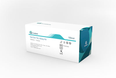
Endotoxin Detection Using Gel-Clot Assay Reagents

,文章长度1000字左右。
# Endotoxin Detection Using Gel-Clot Assay Reagents
## Introduction to Gel-Clot Endotoxin Reagents
The gel-clot method is one of the oldest and most reliable techniques for endotoxin detection. Gel-clot endotoxin reagents play a crucial role in this process, providing a simple yet effective means of identifying bacterial endotoxins in pharmaceutical products, medical devices, and other materials where pyrogen-free status is essential.
## Understanding the Gel-Clot Assay Principle
The gel-clot assay relies on the reaction between endotoxins and a lysate derived from the blood cells of the horseshoe crab (Limulus polyphemus or Tachypleus tridentatus). When endotoxins are present, they trigger a cascade of enzymatic reactions in the lysate, ultimately leading to the formation of a gel clot.
This biological response forms the basis of the test:
1. Sample preparation and dilution
2. Mixing with gel-clot endotoxin reagents
3. Incubation at a controlled temperature (typically 37°C ± 1°C)
4. Visual inspection for clot formation
## Components of Gel-Clot Endotoxin Reagents
### Limulus Amebocyte Lysate (LAL)
The primary component of gel-clot reagents is LAL, which contains the coagulation factors necessary for the reaction with endotoxins. The quality and sensitivity of LAL directly affect the performance of the assay.
### Buffer Solutions
Proper buffering is essential to maintain the optimal pH for the enzymatic reactions. Gel-clot reagents typically include:
– Tris buffer
– Magnesium ions (as cofactors)
– Other stabilizing agents
### Control Standard Endotoxin (CSE)
CSE is used to validate the sensitivity of each batch of reagents and to confirm proper test conditions. It’s derived from Escherichia coli and standardized against the international endotoxin standard.
## Performing the Gel-Clot Assay
### Sample Preparation
Proper sample preparation is critical for accurate results:
1. Selection of appropriate dilution factors
2. Removal of potential interfering substances
3. Confirmation of pH within the acceptable range (6.0-8.0)
### Test Procedure
The standard procedure involves:
1. Reconstituting the lyophilized LAL reagent with endotoxin-free water
2. Preparing serial dilutions of the sample
3. Mixing equal volumes of sample and reagent in depyrogenated tubes
4. Incubating at 37°C for 60 minutes
5. Carefully inverting the tubes to check for clot formation
### Interpretation of Results
A positive result is indicated by the formation of a firm gel that remains in the bottom of the tube when inverted. A negative result shows no clot formation, with the solution flowing freely when inverted.
## Advantages of Gel-Clot Endotoxin Reagents
### Simplicity and Reliability
The gel-clot method offers several benefits:
– Direct visual interpretation without complex instrumentation
– High specificity for endotoxins
– Long history of proven reliability in pharmaceutical testing
### Cost-Effectiveness
Compared to other endotoxin detection methods:
– Lower equipment requirements
– Minimal training needed for technicians
– Reagents are generally less expensive than those for chromogenic or turbidimetric methods
### Regulatory Acceptance
The gel-clot method is:
– Recognized in all major pharmacopeias (USP, EP, JP)
– Accepted by regulatory agencies worldwide
– Suitable for both routine testing and validation studies
## Limitations and Considerations
### Sensitivity Range
Gel-clot reagents typically offer sensitivities ranging from 0.03 EU/mL to 0.25 EU/mL. While adequate for most applications, some situations may require more sensitive methods.
### Subjectivity in Interpretation
The visual endpoint determination introduces some subjectivity, which can be mitigated by:
– Proper technician training
Keyword: Gel-Clot Endotoxin Reagents
– Use of positive controls
– Implementation of standardized interpretation criteria
### Interference Factors
Certain sample characteristics

